Antigen Presentation Complex - The Body's Most Diverse Surveillance System

Imagine that a swarm of T cells, the “police” cells of the immune system, after receiving a signal that the body is being invaded by a virus, has to search for an area containing virus-infected cells mixed with other healthy cells. The job of this T cell is not simply to destroy the whole area of ​​tissue containing a mixed cell population, but to kill the virus-infected cells, leaving the other healthy cells alone. So how does it identify which cells to destroy? In this article, we will refer to antigens as the “identification papers” of cells, and the antigen-presenting complex as the way cells present their “identification papers” to T cells. In addition, other similar functions of the antigen-presenting complex are also mentioned.

1. Antigens and antigen presentation complexes

Antigen is a concept that has been mentioned a lot recently, when the COVID-19 epidemic is a challenge for many countries. Therefore, the demand for rapid testing for COVID is increasing. In fact, some COVID-19 rapid test kit methods are often mentioned as operating based on the principle of recognizing COVID antigens – because antigens are often considered as biological markers for COVID-19. However, the role of antigens in the human body is a biological knowledge that is little known to the public, and even less known about our body recognizing antigens through antigen-presenting complexes. First, we will learn what antigens are and from there, we will go to the definition of antigen-presenting complexes. Understanding the role of this complex helps us to understand how the body fights and recognizes virus-infected cells.

1.1. Kháng nguyên

Antigen is a general term used to refer to all protein, peptide, or polysaccharide molecules originating from both inside and outside the human body. For human antigens, this is simply a protein or polysaccharide originating from within the cell, brought out of the cell wall, and retained by the Major histocompatibility complex Class I ((Major histocompatibility complex Class I), which we will discuss later. This is one of two types of complexes located on the cell wall.

For foreign antigens, its presence inside the body mobilizes the immune system to recognize and attack it. In the context of COVID-19, the antigen can be the spike protein on the surface of the virus (Figure 1)

Figure 1: COVID-19 spike protein structure

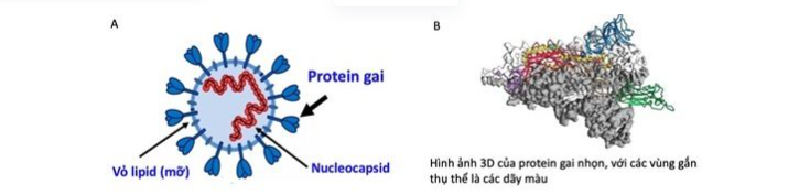
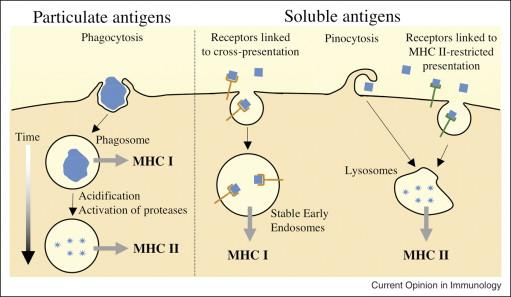
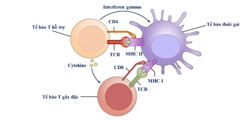
Antigens also play a big role in allergies, where the antigen molecule can be pollen, cat dander, or simply a small particle of dust in the air. Once antigen molecules are present in the human body, they are encountered and “swallowed” by a group of professional presenting cells (Antigen Presenting Cells or APCs) such as dendritic cells and macrophages. Inside these professional enemy recognition cells, they are cut, processed, and pushed out of the cell wall, ready for presentation (Figure 2). Remember that what is presented is not necessarily the antigen, but rather a “shortened” cut version of the antigen – the fragment – and it is the type II antigen presenting complex, which is located on the surface of the cell, that holds this peptide.

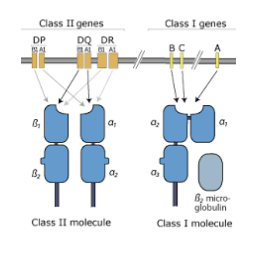
In short, each type of complex will take on the role of a different type of antigen.

            So why do these viral peptides need to be presented, and what does presentation mean?


APCs can be understood as professional “teacher” and “signal” cells. Once they find foreign antigens in the body, they move to the lymph nodes to seek help from T cells – which are simply understood as immune cells that have the role of destroying virus-infected cells. Once there is an alarm signal that the body is being invaded, T cells gather and wait for APCs to train to recognize the enemy. The process of APCs connecting to T cells through the presentation complex (Figure) is called antigen presentation.

1.2. Antigen presentation complex

So what is the structure and mechanism of MHC for antigen presentation? In the body, there are many different types of helper T cells created by the VDJ recombination process, but not all types are suitable for destroying a certain type of “enemy”. Therefore, the antigen presentation process is both meaningful in declaring the new invader of the body and meaningful in selecting the best weapon to destroy the enemy. In this section, we will explain in more detail the mechanism of the presentation process and the structure of MHC, specifically MHC I.

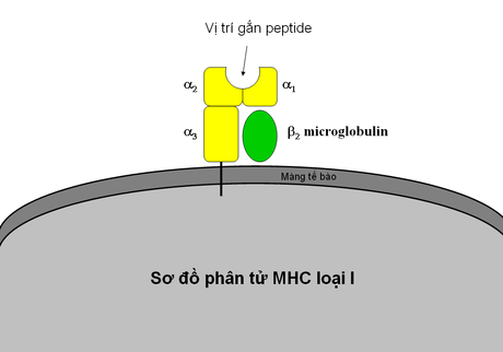
The MHC class I complex has 4 main parts (alpha 1,2,3, and Beta 2). The shape and properties of alpha 2 and alpha 1 will shape the peptide binding site – the edited fragment of the antigen.

Here, we consider an example that includes dendritic cells (a type of APC), helper T cells, and cytotoxic T cells. Dendritic cells carry MHC II with antigen fragments attached, and it binds to helper cells through the T cell receptor. The TCR can be considered the eyes and ears of the T cell, the bridge to receive information from external factors. Of the countless T cells in the body’s reserve, only T cells with grooves that “match” the MHC II – fragment combination can bind to dendritic cells. These “suitable” T cells will multiply in the lymph nodes, and they will be responsible for destroying diseased cells.

The presentation between APC cells via MHC II is only half of the picture of the presentation complex. In addition to MHC II, MHC I is equally important. Let’s imagine that after receiving a signal about the invasion of a virus, the APC cell also signals the T cell, and the cytotoxic T cell rushes to find the target – the virus-infected cell – to destroy. MHC I was probably born from the need to distinguish between enemy and friend for cytotoxic T cells. With healthy cells, they will have fragments of the body’s antigen attached to MHC I, but infected cells will have fragments of the virus’ antigen, because they have been possessed by the virus to become a replication factory. MHC I functions like a student card or an ID card, used to present to the security guard. Remember that this card is only used when the security guard requests it, just as T cells only check when there are signs of invasion. This screening helps the cytotoxic T cells not to kill healthy cells indiscriminately, but to focus on the infected cells only.

Through this, we have understood the mechanism by which the body’s defense cells can talk to each other through the presentation complex II, just as normal cells and defense cells talk through the presentation complex I.

2.Polymorphism of the antigen presentation complex

Above we have briefly discussed the role and structure of MHC in the human body. In this section, we will continue to learn about the diversity of MHC. In the human community, each type of MHC I has countless variations, and their ability to bind to peptides is extremely diverse. This diversity will affect its ability to adhere to T cells, as well as its ability to capture antigens. MHC itself is a cellular complex, produced from genes, and its properties will depend largely on the genes that make it up.

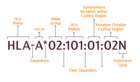
In order to serve the purpose of binding to a wide variety of antigens, MHC is famous in biology for its polymorphism. IMGT/HLA Website ( ) is the largest database currently available that compiles statistics on HLA diversity, as well as storing detailed genomes for each specific complex. There are 30,522 different HLA complexes, representing only 45 genes and pseudogenes. To systematize such a huge number, a clear nomenclature system is needed. It should be noted that this nomenclature is only nominal, or a convenient way of classifying.

Each genotype of an HLA gene is called an allele. An allele is usually identified by a hierarchical system. Initially, the group of alleles will be denoted by the gene name, such as A, B, C if they belong to MHC I, or DRB1, DQB1, etc. if they belong to MHC II. The next 4 levels, separated by the “:” sign, will gradually go into the details of the allele. Just as an allele can be considered a student of a school, that student can be a person of a group, a group of a class, a class of a grade, etc. Therefore, we can imagine the huge number of “MHC schools”, when each allele needs 5 such levels of classification.

The more levels an allele is described by, the more details we know about it. For example, when we look at the allele name HLA-A*02:101, we know its protein sequence, but with HLA-A*02:101:01 we also know the gene sequence that codes for this protein, and with HLA-A*02:101:01:01, we even know the entire HLA-A gene sequence, including the non-coding segments.

The polymorphism of the MHC group alleles, of course, also causes many headaches for scientists. Typically, with a linear reference Genome (link to this article), it is impossible to have a sequence representing more than a combination of 30 thousand alleles, so the results of gene sequence alignment on the MHC gene segment are often very unreliable. Therefore, specialized tools are needed to accurately determine the MHC combination in each person’s genome. Currently, the 1000 Vietnamese Genomes Project has mastered the analysis process to determine MHC I and II, and is publishing results with high reliability, verified by many data sources here.  VinGen Data Portal (vinbigdata.org).

In the next series of articles, we will address the question: do MHC polymorphisms affect disease resistance, and how? How to design experiments to validate the results of MHC analysis?

Leave a Reply

Your email address will not be published. Required fields are marked *